
We specialize in high-risk pregnancy.
We work to minimize complications and help you achieve the healthiest pregnancy possible.
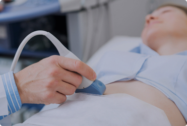
It’s used throughout pregnancy to monitor your baby’s growth, detect potential concerns early, and evaluate gynecologic conditions when symptoms arise. With standard 2D imaging alongside advanced 3D and 4D technology, our team delivers a level of detail that keeps you informed and your care on solid footing.
Understanding Pregnancy Ultrasound Services
Ultrasound works through the use of high-frequency sound waves that travel through the body and reflect off internal structures. These reflected waves send information back to a computer, which translates the data into clear images displayed on a screen. This process does not involve radiation, which makes ultrasound safe to use at any point during pregnancy.
Two primary methods are used depending on the patient’s needs and stage of pregnancy. A transabdominal ultrasound uses a handheld transducer moved across the abdomen to capture images of the uterus and baby. A transvaginal ultrasound uses a slender transducer placed inside the vagina, which produces more detailed images because of its closer proximity to pelvic organs.
Physicians carefully choose the most appropriate method based on each individual situation.
What to Expect During a Pregnancy Ultrasound
First-trimester ultrasounds are not always routine, but our physicians may recommend one to verify a pregnancy, confirm its location, or establish an estimated due date. A second-trimester ultrasound is standard for all pregnant patients and typically occurs between 18 and 20 weeks.
This scan evaluates the baby’s size, position, anatomy, and amniotic fluid levels. After twenty weeks, the external genitalia often become visible, which gives many parents the opportunity to learn the baby’s sex. Some situations may limit visibility if the baby’s position blocks the view during the scan.
Advanced 3D and 4D Ultrasound Technology
At Rosh MFM, we offer state-of-the-art 3D and 4D ultrasound imaging as part of our pregnancy ultrasound services. A 3D scan produces a detailed, static image of your baby’s facial features and anatomy with remarkable precision. A 4D scan captures live, streaming video of your baby in motion, including yawning, stretching, and heart-valve movement.
These advanced scans are available from 27 weeks onward and serve both diagnostic and personal purposes. Many parents request 3D and 4D imaging to experience a vivid, moving look at their baby before birth. Our physicians use the same technology to assess cardiovascular structures and fetal anatomy in high-resolution detail.
Ultrasound for High-Risk Pregnancy Monitoring
Patients with high-risk pregnancies receive more frequent and detailed ultrasound monitoring throughout their care. Our maternal-fetal medicine specialists use advanced imaging to track fetal growth, assess placental health, monitor amniotic fluid levels, and evaluate structural concerns as they develop. Ultrasound also plays a key role in guiding procedures such as amniocentesis and cordocentesis.
Beyond pregnancy, ultrasound helps our physicians diagnose a range of gynecologic conditions by producing detailed images of the uterus, ovaries, and fallopian tubes. Common conditions identified through ultrasound include endometriosis, uterine fibroids, ovarian cysts, and ectopic pregnancy.
See Your Baby In Extraordinary Detail
At Rosh MFM, our advanced 2D, 3D, and 4D ultrasound technology gives you and your physicians a remarkable window into your baby’s health and development. Precise imaging at every stage means nothing goes unnoticed.
Recognition and Credentials
Dr. Daniel F. Roshan, MD, FACOG, FACS, holds dual board certifications in Obstetrics and Gynecology and Maternal-Fetal Medicine.
Rosh MFM is recognized across New York for its advanced imaging capabilities, including one of the most experienced 3D and 4D ultrasound programs available in an outpatient OB/GYN setting. Our certified genetic counselor adds another layer of expertise to help patients with complex prenatal results.
What Our Patients Are Saying
Hearing directly from patients reminds us why investing in advanced technology and attentive care makes such a difference. Here is what a few of our patients have shared:
“I enjoyed my visit. The staff was professional, friendly, and answered all my questions without me even having to ask.” — Deasia J.
“Exceptional doctor!” — Alketa B.
Your baby deserves to be seen clearly, and so do you. Book your ultrasound appointment with our New York team and experience imaging that goes beyond the standard.
Contact Us
Disclaimer: The content on this page is intended for general informational purposes only and does not constitute medical advice. Always consult your healthcare provider before making any decisions about your health or pregnancy care.
Ultrasound uses high-frequency sound waves to create images of organs and structures inside your body. As sound waves travel through your body, they reflect off tissues, sending information to a computer that interprets them and produces an image.
Your ultrasound may be performed transabdominally or transvaginally. A transabdominal ultrasound is the type used for pregnant women. The devices that emit sound waves, called transducers, are moved across the abdomen, sending the waves toward the uterus.
A transvaginal ultrasound uses a long, narrow transducer that’s placed inside the vagina. This method produces a clearer image of your pelvic organs because it’s closer to them.
Ultrasound is safe to use during pregnancy to evaluate the baby’s growth and development because it relies on sound waves rather than radiation at any time during pregnancy.
First-trimester ultrasounds aren’t routine, but they may verify a pregnancy, determine its location, and estimate the due date. Every pregnant woman receives a second-trimester ultrasound to determine the baby’s size and position.
Your baby’s sex can often be identified when the second-trimester ultrasound is done after 20 weeks because the external genitalia are visible. In some cases, however, the image may not be clear, or the baby’s position may block the view.
The team at Rosh Maternal & Fetal Medicine offer state-of-the-art 3D and 4D ultrasounds. With 3D imaging, you can see the facial structure and anatomy as a static three-dimensional image. With 4D ultrasound, live streaming video of the real-time images is captured, showing movement like the baby yawning or the motion of heart valves.
Ultrasound can help diagnose many different reproductive health problems as it shows detailed images of your uterus, ovaries, and fallopian tubes. Your doctor may use ultrasound to determine the cause of pelvic pain and abnormal bleeding.
A few of the most common conditions diagnosed with ultrasound include:
Come visit your Manhattan OBGYN.
We work to minimize complications and help you achieve the healthiest pregnancy possible.